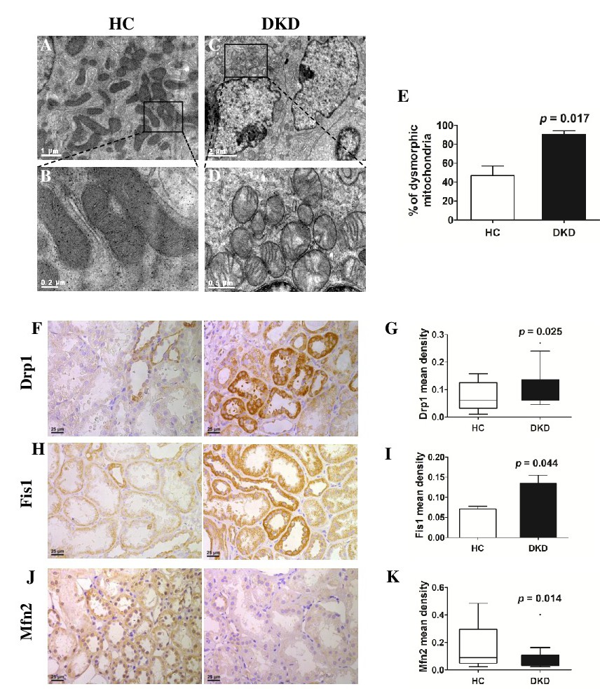
Fig. 2. Mitochondria were fragmented in proximal tubules of human kidney biopsy tissues from DKDs. (A-D) By electron microscopy, mitochondria fragmented into short rods or spheres in DKDs (n=3), and the elongated mitochondria with organized cristae observed in HCs (n=3). (E) The mitochondrial changes in electron micrographs were quantified and included in bar graphs. (F-K) The mitochondrial fission protein Drp1 and Fis1 was notably increased and the fusion protein Mfn2 was decreased in renal tubules compared to HCs. ×400 magnification. HC, healthy control; DKD, diabetic kidney disease.